Researchers have developed a digital solution using AI technology and a mobile application to significantly speed-up skin cancer diagnosis.
Skin cancer is a particularly deceptive form of cancer. In the early stages, it visually looks like a harmless mole or birthmark and causes no pain. According to data from the German Cancer Society, more than 200,000 people suffer from skin cancer every year in Germany. The earlier a patient has a skin cancer diagnosis, the more treatable it is.
The Fraunhofer Center for Assistive Information and Communication Solutions (AICOS) in Porto and Lisbon has developed a solution to accelerate early recognition. The Derm.AI solution combines smartphone photos of the skin lesion with image-analysis software and artificial intelligence. It provides a swift, first assessment of potentially dangerous changes in the surface of the skin. Dermatologists can access this decision support platform and analyse the cases with increased risk of skin cancer first. The Derm.AI solution aims to improve the existing Teledermatology processes in the Portuguese National Health System.
“In recent years, GPs have been increasingly concerned with spotting skin cancer early. People who notice dark spots or other perceptible changes to their skin need clarity quickly. But in regions with few specialists, it often takes a long time to get an appointment for the initial assessment. Often patients also have to travel long distances for these appointments too. This is where our Derm.AI solution comes in,” said Maria Vasconcelos, Senior Scientist at Fraunhofer AICOS.
Accessible smartphone app
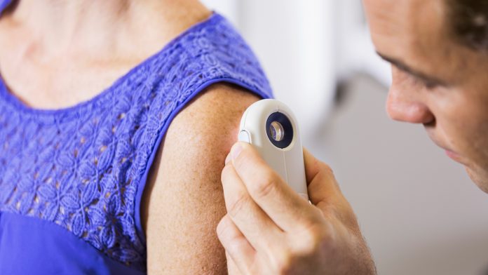
The Fraunhofer team have developed a specialised app for GPs to photograph the potential skin cancer area with a smartphone – it will be available for both iPhones and Android smartphones. The app ensures the photos are correctly aligned, taken at the right distance and have the correct resolution. The app takes two photos: one close-up of the area in question and another image further away to show the area in context. It also helps to correctly align and position the camera. This creates standardised photos with consistent settings for resolution, colour, brightness and contrast.
“The standardised shots are easy to compare and can be reliably analysed by specialists,” Vasconcelos explained.
AI technology
The images obtained at the GP practice are sent to the dermatology department of a hospital. AI software will analyse the images of the skin lesion, compare them to reference data and data of other patients then provides a risk assessment. The lesion in question is labelled as “normal”, “priority”, or “high priority.” This is not a formal skin cancer diagnosis; it is a first assessment that helps prioritise the order in which the potential cases are examined. The doctors can prioritise examining the cases which the AI software has indicated have a high risk, as these need to be quickly confirmed or clarified.
“The software doesn’t make a decision; it simply provides a pre-selection based on probability. The actual examination and diagnosis are still in the hands of the dermatologists or skin-cancer specialists,” Vasconcelos explained.
After the images are analysed, along with the patient data such as age, gender or previous conditions, the dermatologist at the hospital can either provide feedback to the GP responsible for the patient via teleconsultation or schedule a presential consultation with the patient.
During the Derm.AI project, the AICOS researcher and her team developed the algorithm for the image-analysis software. The deep-learning software was fed with image data and information from around 4000 cases. The algorithm also utilised the expert knowledge of dermatologists in the subsequent prioritisation.
“We had a lot of discussions with GP and dermatologists to understand what they need. We have received very good feedback from doctors for Derm.AI,” Vasconcelos stated.
Skin Cancer diagnosis
A dermatologist will book an in-person appointment if there is some uncertainty surrounding the risk level of the skin lesion. An examination of the skin lesion under a reflected-light microscope will take place, or a tissue sample for analysis will be carried out, which will allow the specialist to accurately deliver a skin cancer diagnosis.
Around 80% of the cases in which patients present themselves at the GP’s practice with a suspicious change in their skin prove to be harmless moles or birthmarks, after image analysis and consultation between the doctor and the dermatologist. This enables GPs to quickly give patients the all-clear, saving them long periods of waiting and an often-long journey to a hospital appointment.
However, the new digital solution speeds the process of getting patients an accurate skin cancer diagnosis and allows prioritisation of cases to ensure high-risk cases are being addressed first.
For patients whose skin changes are not identifiable as harmless or for those which indicate a less dangerous form of skin cancer, the GP asks the patient to return, for example, in three months and have another photo taken of the area.


