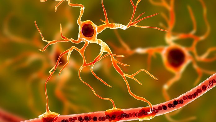
A Weill Cornell Medicine study has identified that brain cells called astrocytes may be influential in causing symptoms of autism spectrum disorder.
The Weill Cornell Medicine researchers developed astrocytes from the stem cells of autism patients and transplanted them into healthy newborn mice, findings that abnormalities in these brain cells may be a cause of autism spectrum disorder symptoms.
The researchers observed that the mice developed repetitive behaviours after the transplant, a characteristic symptom of autism, but they did not display the social deficits of the disease. Additionally, the mice developed memory deficits that are common in autism spectrum disorder but not a primary characteristic of the condition.
Dr Dilek Colak, an assistant professor of neuroscience at the Feil Family Brain and Mind Research Institute at Weill Cornell Medicine, commented: “Our study suggests that astrocyte abnormalities might contribute to the onset and progression of autism spectrum disorders. Astrocyte abnormalities may be responsible for repetitive behaviour or memory deficits, but not other symptoms like difficulties with social interactions.”
What do astrocytes do?
Previous research into autism spectrum disorders has primarily analysed the impact of neurons, brain cells that transport information throughout the brain. However, the importance of astrocytes, which are essential in moderating the behaviour of neurons and the connections between them, is relatively unstudied territory.
Genetic mutations associated with autism spectrum disorder potentially impact a range of brain cells in different ways, with post-mortem research identifying abnormalities in the brain of patients with the disease.
Dr Colak said: “We didn’t know if these astrocyte abnormalities contributed to the development of the disease or if the abnormalities are the result of the disease.”
How do they affect autism spectrum disorder?
For their astrocyte investigation, the team acquired stem cells from autism patients and stimulated them into creating astrocytes, which they then transplanted into the brains of healthy newborn mice, resulting in a human-mouse chimaera. Subsequently, the team employed two-photon imaging to examine excessive calcium signalling in the transplanted human astrocytes.
Dr Ben Huang, instructor of neuroscience in psychiatry at Weill Cornell Medicine, said: “It was amazing to see these human astrocytes responding to behavioural changes in active mice. We believe we are the first to record the activity of transplanted human astrocytes this way.”
Next, the team looked to determine if this elevated calcium signalling was causing the behavioural symptoms in the mice. To do this, the team infected astrocytes from autism patient stem cells with a virus that included a fragment of RNA designed to mitigate calcium signalling to normal levels. When these astrocytes were transplanted, the mice did not develop memory problems.
“Future therapies for autism might exploit this finding by using genetic tools to limit extreme calcium fluctuations inside astrocytes,” said co-lead author Megan Allen, a postdoctoral associate in neuroscience in the Feil Family Brain and Mind Research Institute at Weill Cornell Medicine.
The team stated that their findings might be essential for understanding the mechanisms behind other neuropsychiatric diseases, such as schizophrenia.
Dr Colak concluded: “It is important to determine the roles of specific types of brain cells, including astrocytes, in neurodevelopmental and neuropsychiatric diseases.”


