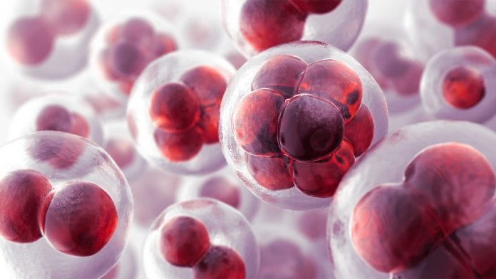
When cells are stressed, they initiate a complex and precisely regulated response to prevent permanent damage.
Researchers from Goethe University have developed a new protoeomics procedure to help measure acute cellular changes. This can help us understand the role of stressed cells in the development of disease; how underlying molecular processes work.
The new method opens the door for the development of new therapeutic strategies.
One of the immediate reactions to stress signals is a reduction of protein synthesis. Before this research, published in Molecular Cell, it was difficult to measure such acute cellular changes.
Measuring cellular changes
The team, led by biochemist Dr Christian Münch, who heads an Emmy Noether Group, employs a simple but extremely effective trick: when measuring all proteins in the mass spectrometer, a booster channel is added to specifically enhance the signal of newly synthesised proteins to enable their measurement.
Thus, acute changes in protein synthesis can now be tracked by state-of-the-art quantitative mass spectrometry.
The idea emerged because the team wanted to understand how specific stress signals influence protein synthesis.
Group leader Münch, said: “Since the amount of newly produced proteins within a brief time interval is rather small, the challenge was to record minute changes of very small percentages for each individual protein.”
The newly developed analysis method now provides the team with detailed insight into the molecular events that ensure survival of stressed cells.
Cells play a vital role in the development of disease
The cellular response to stress plays an important role in the pathogenesis of many human diseases, including cancer and neurodegenerative diseases.
An understanding of the underlying molecular processes opens the door for the development of new therapeutic strategies.
Münch said: “The method we developed enables highly precise time-resolved measurements. We can now analyse acute cellular stress responses, i.e, those taking place within minutes. In addition, our method requires little material and is extremely cost-efficient.
“This helps us to quantify thousands of proteins simultaneously in defined time spans after a specific stress treatment.”
Due to the small amount of material required, measurements can also be carried out in patient tissue samples, facilitating collaborations with clinicians.
At a conference on Proteostatis in Portugal, PhD student Kevin Klann was recently awarded with a FEBS journal poster prize for his presentation of the first data produced using the new method. The young molecular biologist demonstrated for the first time that two of the most important cellular signalling pathways, which are triggered by completely different stress stimuli, ultimately results in the same effects on protein synthesis.
This discovery is a breakthrough in the field.


