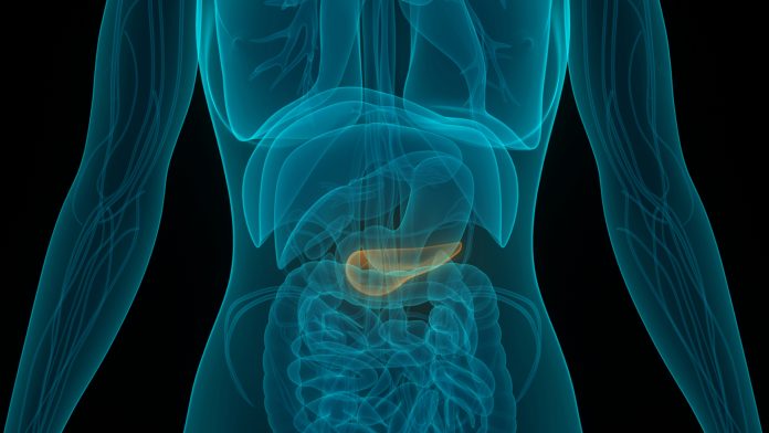
A study from the Systems Oncology Lab at Champalimaud Research, published on September 9, has provided a potential solution to the issues surrounding pancreatic cancer treatment. The study explores the use of extracellular vesicles (EVs) in fighting cancer.
Statistics surrounding pancreatic cancer are not positive. The five-year survival rate after a pancreatic cancer diagnosis is just 9% and pancreatic ductal adenocarcinoma (PDAC), the most common form of pancreatic cancer, is projected to be the second highest cause of cancer deaths by 2030.
Currently, surgery is the most effective pancreatic cancer treatment, but for 70-80% of patients, surgery is not a viable option. It is widely understood that understanding cancer at the cellular and subcellular level is essential for developing better pancreatic cancer treatment.
Pancreatic cancer treatment typically includes a mix of radiation and chemotherapy. Presently, assessing the progress of PDAC patients after therapy relies on imaging and measuring blood serum levels of cancer biomarkers.
However, both these methods are flawed. Imaging (CT, MRI) cannot detect small tumours or differentiate between benign and malignant tumours. Even the most well-established PDAC marker is not present at 5-20% in PDAC patients and can lead to unreliable results.
Using EVs in pancreatic cancer treatment
“EVs are tiny sacs released by cells. They are like mini-cells, with a lipid membrane, genetic material, proteins and sugars. For a long time, they were dismissed as ‘trash bags’, filled with waste products that cells wanted to throw out. But it’s now clear that they also transfer messages between cells,” explained Bruno Costa-Silva, principal investigator, and the study’s senior author.
It is believed by the researchers, that EVs may be one of the most powerful forms of communication in living organisms, as they are also released by plants and bacteria. EVs are produced by virtually all cells, including cancerous ones. Studies have even found that EVs can significantly contribute to tumour progression.
Previous research by Costa-Silva has found that EVs in the blood could be used to detect, predict, and locate pancreatic cancer metastases. “Ours is the first study to show that by looking at how EVs in pancreatic cancer patients change over time, we can tell how well they are responding to therapy”, said Costa-Silva.
Surprising results found during research
When the researchers began investigating the potential of EVs for monitoring pancreatic cancer treatment response, they found surprising results. “At first, we thought it was an artefact, but it soon became clear that EVs in PDAC patients had significantly higher levels of a specific protein compared to EVs in healthy controls” said Nuno Couto, oncologist at the Champalimaud Foundation.
The protein in question was Immunoglobulin G (IgG), a type of molecule that is part of the body’s defence system, which finds and kills cancer cells.
To see how levels of these IgG-positive EVs changed in patients during pancreatic cancer treatment, the researchers had to collect 20-30 blood samples from the same patient over a series of months.”
The researchers rapidly measured EV populations in blood samples. They found that IgG-positive EVs increase during disease progression and decrease in response to pancreatic cancer treatment. Subsequently, the EVs represent a new biomarker that will add to the repertoire of tools available to evaluate a tumour’s status.
“We were very excited to see such a close correlation between these vesicles and therapy response”, said Costa-Silva. “We now have a more reliable tool to assess and improve, the efficacy of PDAC treatments, and to reduce the unnecessary and harmful side effects of ineffective ones. It’s impossible to look at these results, and not to think of immunology, or the broader implications for cell signalling”.
The researchers discovered that IgG binds to EVs in PDAC patients via a cancer antigen. It is suspected that EVs expressing this antigen are released by the tumour itself. IgGs become bound to the EVs instead of to their intended target: the tumour cells. This way, the tumour can evade the immune system and launch EVs to intercept IgGs.
“If very aggressive cancers like PDAC use EVs to disarm the immune system, we can develop new therapies to target tumour-derived EVs and make these cancers less resistant to treatment,” concluded Costa-Silva.


