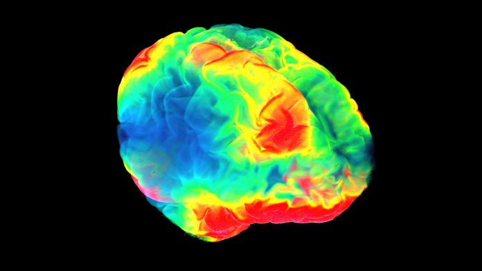
Researchers at Karolinska Institutet in Sweden are using novel 3D imaging technology to better understand the early stages of Alzheimer’s disease.
Using new 3D imaging technology, researchers have been able to comprehensively characterise the part of the brain that may show the earliest accumulation of tau protein, an important biomarker in the early stages of Alzheimer’s. The results may enable doctors to make more precise neuropathological diagnoses of the Alzheimer’s disease spectrum in the early stages of the disease.
Intracellular accumulation of pathological tau protein in the brain is a key indicator of several age-related neurodegenerative disorders, including Alzheimer’s disease, which accounts for 60%-80% of all dementia cases.
New discoveries made using 3D imaging
The researchers at Karolinska Institutet, SciLifeLab in Stockholm worked alongside universities in Hungary, Canada, Germany, and France. They used state-of-the-art volume immuno-imaging technology, to investigate a human brainstem nucleus called locus coeruleus, which is a key hub in the mammalian brain.
Locus coeruleus is a small brain nucleus and is very hard to study using existing 2D imaging methods. When the locus coeruleus was analysed using 3D imaging, researchers found it had an intriguing complexity. They found that the locus coeruleus had previously undescribed cellular forms of tau pathology in the brain region during the early stages of Alzheimer’s disease.
“Our study shows that a gradual dendritic atrophy is the first morphological sign of the degeneration of tau-bearing locus coeruleus neurons, even preceding axonal lesion,” said the study’s last author Csaba Adori, a researcher at the Department of Neuroscience, Karolinska Institutet.
“Dendrites are crucial nerve fibres through which the neurons communicate, and dendritic degeneration leads to functional deficits like hyperactivation of neurons. This may contribute to several symptoms that precede the onset of Alzheimer’s disease, like sleep disturbances, anxiety, and depression, which are consistent with locus coeruleus dysfunction,” explained Adori.
New technology may help to treat the early stages of Alzheimer’s
The 3D imaging technology revealed a non-random distribution of tau-bearing locus coeruleus neurons with a clustering tendency. The researchers also found the dendrites of adjacent, clustered tau-bearing neurons frequently showed dendro-dendritic contacts. This discovery supported the theory that pathological tau may spread via neuronal processes from one neuron to the other, this idea had previously been suggested by in vitro studies and animal experiments.
As well as this, the researchers were able to demonstrate that tau pathology is more prominent in the part of the locus coeruleus that projects to forebrain regions that are heavily affected in the early stages of Alzheimer’s disease.
These results may represent a significant advance in the scientist’s understanding of how brain cell architecture may have an impact on the development and spreading of pathological tau protein in the early stages of Alzheimer’s.
“Our results help to further understand why certain brain regions are more affected in Alzheimer’s disease than others,” said Adori. “Moreover, the novel methodical approach applied to human tissue opens the way for more precise neuropathological diagnostic procedures, even in very early stages of the Alzheimer’s disease spectrum. This hopefully contributes to working out more effective prevention strategies in the future.”
The results of the study were published in the journal Acta Neuropathologica.


