New research has found that an imaging process used in research labs could detect early-stage lung disease if developed for use in hospitals and clinics.
Researchers from the KTH Royal Institute of Technology in Stockholm examined how a process called phase-contrast X-ray imaging could be used to detect lung disease in humans. In the study titled ‘Phase-contrast virtual chest radiography‘, the research team used a model developed at Duke University that simulates the human chest.
The researchers found that phase-contrast chest radiography could visualise the smallest airways in the lungs, including those less than 2mm wide. The technology can also help to identify lung disease-related obstructions, something that conventional radiography cannot do.
The limitations of conventional imaging
Conventional Phase-contrast imaging used in research labs is limited to imaging centimetre-scale samples of soft tissue. This study has shown that phase-contrast X-ray imaging has the potential to detect lung disease if the technical demands can be engineered for clinical use.
According to the study’s lead author, Ilian Häggmark, current chest radiography methods used in clinics and hospitals today can play a role in detecting lung disease, but it is limited by how it generates images.
“Phase-contrast X-ray imaging can extract more information at higher resolution using the same amount of radiation dose as in conventional radiography,” said Häggmark.
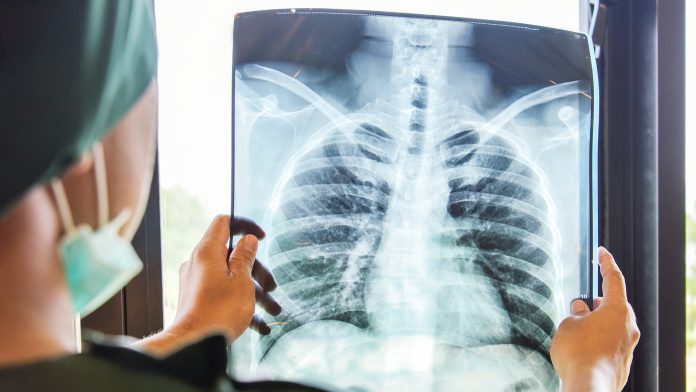
In conventional radiography, the X-ray beam passes through the body before it is absorbed by different tissues in different amounts. A detector then measures the beam’s intensity after filtering through the body. This process is known as attenuation and is the basic mechanism for providing the contrast that forms X-ray images.
The phase-contrast technique allowed clinicians to extract more information from each X-ray beam. Using this technology, clinicians can measure differences in the waveforms of X-rays that pass through a sample. X-ray beams encounter atoms and other structures that can change the position of a wave at any point in relation to a reference wave.
Phase-contrast technology makes it easier to detect lung disease
This information is used to generate an image that enhances structures in the sample. These images can highlight the boundaries of bronchial walls and small airways with higher contrast and better resolution, enabling clinicians to easily identify signs of lung disease.
The researchers have said that moving the detector further away from the patient is key to this method. They also emphasise that developing equipment for imaging larger samples will take time.
“You need an X-ray source with both high power and a small emission spot. Basically, you need bright X-ray sources,” said Häggmark.
According to the researchers, promising developments are taking place, but it will take time for these to be ready for human use.
“For now, simulations and virtual clinical trials are the perfect tools to explore what we can do when the source technology is ready,” concluded Häggmark.


