Dr Sarah Hardcastle, Consultant Rheumatologist at the Royal National Hospital for Rheumatic Diseases, spoke to Health Europa about pregnancy-associated osteoporosis.
When we think of osteoporosis, we often associate the condition with older members of our society, rarely expecting pregnant women to be affected. Indeed, research around what could cause bone fractures during pregnancy is still very much in its infancy, with many cases going undiagnosed or prompting further investigation due to the rarity of the condition. Pregnancy-associated osteoporosis (PAO) usually occurs during late pregnancy or early postpartum, most commonly manifesting as bone breakage in the spine.
Dr Sarah Hardcastle is a Consultant Rheumatologist at the Royal National Hospital for Rheumatic Diseases who, in 2019, published a paper which detailed a review of pregnancy-associated osteoporosis cases at the hospital in which she works. Here, she speaks to Health Europa about potential risk factors, symptoms and treatments.
How common is pregnancy associated osteoporosis (PAO)?
Pregnancy-associated osteoporosis is undoubtedly very rare, but it is difficult to be precise about the numbers of patients affected. This is because the condition has not been systematically studied, and the relative contribution of pregnancy versus other risk factors for fracture in individual women with PAO is not always clear. It is also likely that pregnancy-associated osteoporosis is underdiagnosed, as many women who fracture during or after pregnancy may not be referred for further investigations such as bone density scans.
What causes pregnancy associated osteoporosis?
The cause of pregnancy-associated osteoporosis is not fully understood and is still being researched. A proportion of women who fracture during or after pregnancy turn out, on further investigation, to have additional risk factors such as low vitamin D levels, smoking or previous use of medications such as steroids which are known to increase fracture risk. A very small proportion may have an underlying genetic bone disorder (such as osteogenesis imperfecta) which was previously undiagnosed. However, in the great majority of women with PAO, a cause cannot be identified. Bone biopsy studies have suggested that the osteoblasts (bone-forming cells) of women with PAO may be underactive, but these studies have been small and further research is needed.
What are some common signs and symptoms of PAO? When do fractures usually occur?
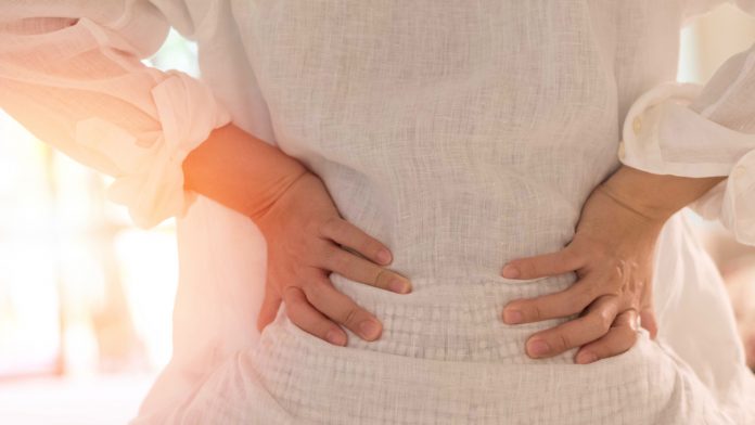
The first sign of pregnancy-associated osteoporosis is usually the onset of acute back pain in the latter stages of pregnancy or shortly after delivery. This is because vertebral (backbone) fractures are by far the most common fracture type in this condition. Affected women may also notice height loss, or a change in the shape of their back. The reason vertebral fractures occur predominantly is thought to relate both to the type of bone present in the vertebrae (trabecular or ‘woven’ bone), and the additional strain that pregnancy places upon the spine. Unfortunately, because back pain is perceived as being common both during and after pregnancy, many women are not investigated further for their back pain for several weeks, leading to delays in diagnosis. In PAO, fractures occurring during pregnancy usually happen during the third trimester (last three months of the pregnancy), but the majority of fractures actually occur after delivery, usually around the three-month mark. Most women develop PAO during their first pregnancy, though later pregnancies can sometimes be affected.
What are the key risk factors in PAO? Are there any preventive lifestyle measures that can be taken to lessen the risk of PAO?
The rarity of pregnancy-associated osteoporosis makes it difficult to identify consistent risk factors for the disorder. As mentioned above, we do know that a proportion of women with PAO have additional risk factors for low bone density and fracture such as smoking, low body weight, use of certain medications and low vitamin D levels. A couple of studies have suggested that women with PAO are more likely to have a family history of osteoporosis and / or fractures, so genetic factors may well be important. One study examining risk factors for PAO found that immobilisation during pregnancy as a result of complications like hyperemesis gravidarum or pre-eclampsia was more common in women with PAO compared with a control group, and the same study suggested that affected women may have been less physically active earlier in life. However, a large proportion of cases of PAO remain unexplained. It is likely that the usual bone health lifestyle measures such as maintaining an adequate intake of calcium and vitamin D, regular weight-bearing exercise and avoiding smoking and excessive alcohol consumption may reduce the likelihood of developing PAO. However, we currently have no data to prove that these measures are effective, and they are unlikely to be able to prevent all cases of PAO.
What treatments are currently available for women experiencing PAO?
Treatment usually starts with simple measures such as calcium and vitamin D supplementation. Pain relief and / or physiotherapy can be helpful for painful vertebral fractures. Women who develop PAO are also generally advised to stop breastfeeding, as we know that this can lead to a further decline in bone density which is obviously best avoided in women who have had a recent fragility fracture. However, it is important to emphasise that breastfeeding is safe and recommended for the vast majority of women who do not have PAO, as the declines in bone density seen during lactation are fully reversible once weaning occurs.
In addition to the above measures, registry studies have also shown that a significant proportion of women with PAO receive treatment with traditional osteoporosis medications such as bisphosphonates. However, the evidence that these medications are effective in the context of PAO is very limited. This is partly because there have been few studies in this specific patient group, and also because those studies have generally not included a control group of women who did not receive treatment for the purposes of comparison. One of the challenges of assessing treatment effects in PAO is that bone density generally improves spontaneously within six to 12 months once pregnancy and lactation has been completed. Therefore, without a control group, it is hard to say whether any observed improvements in bone density are as a result of the treatment or would have occurred anyway. Ideally, we need randomised controlled trials to address this question, but it may be difficult to recruit enough patients because pregnancy-associated osteoporosis is so rare. Importantly, most osteoporosis medications are contraindicated during pregnancy itself, so can only be considered after delivery.
Dr Sarah Hardcastle
MBChB, Bsc, MRCP (UK), PhD,
Consultant Rheumatologist
Royal National Hospital for Rheumatic Diseases
www.ruh.nhs.uk
This article is from issue 19 of Health Europa Quarterly. Click here to get your free subscription today.


