
Christian Stockmann, Assistant Professor at the University of Zurich, outlines his recent research into natural killer cells and how these cells can be harnessed to accelerate wound healing, and fight microbial infection.
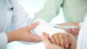
The treatment and management of wounds is an increasingly important problem in Europe, and across the globe. With antimicrobial resistance on the rise, and a greater prevalence of non-communicable diseases (NCDs) worldwide, more and more people are becoming vulnerable to chronic wounds which can take up to eight weeks to heal. There are an estimated 15 million wound patients in the EU,1 and therefore, a necessity to find different ways to advance wound healing treatments. Indeed, today only 30-40% of patients in the EU receive adequate wound therapy.1
Recent research led by Christian Stockmann, Assistant Professor at the University of Zurich, has found that natural killer cells help with the management of wounds. Here, Stockmann tells Health Europa more about his research and discusses how these killer cells could accelerate wound healing.
What are some of the significant challenges facing the management and treatment of wounds today?
Major challenges in the successful treatment of chronic wounds are impaired regenerative ‘healing’ responses such as insufficient angiogenesis and hence, poor perfusion of the wound bed, along with impaired epithelialisation. This is further complicated by an associated risk of wound infection despite unresolved inflammation.
Are there any particular demographics who are more at risk of chronic or difficult-to-treat wounds?
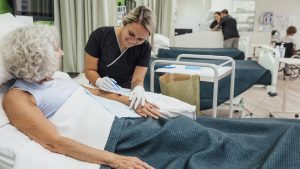
The costs for wound care are a considerable burden for healthcare systems and are projected to further increase, owing to the rising prevalence of diseases (e.g. diabetes, neurodegenerative disorders, and heart disease), which negatively impact on wound healing, along with increasing bacterial resistance to antibiotics.
What can be done to minimise the risk of infection when treating difficult wounds?
Common efforts include the use of antibiotics, the optimisation of surgical techniques, or measures to maintain sterility of the wound bed. However, it is well documented that all wounds become contaminated, often by bacteria from the skin or within the patient. It is the host’s immune system that prevents such contaminations from developing into clinical infections. This host-defense mechanism is particularly important during the initial hours and the first days following contamination.
Can you tell us about your research on natural killer cells?
After tissue injury, an adequate immune and repair response are prerequisites for rapid wound closure and the prevention of microbial infections. Skin injury triggers key components of the skin repair and defence machinery including inflammation and regenerative angiogenesis, and these must be tightly coordinated. Innate lymphoid cells (ILCs) are an early source of cytokines, particularly NKp46+ group 1 ILCs (ILC1s) that comprise natural killer (NK) cells, guide inflammation, and tailor the immune response to the type of the encountered insult. While the role of skin-resident ILC1s for steady-state skin homeostasis is increasingly recognised, the significance of infiltrating NK cells for skin repair and antimicrobial defence remains unknown.
NK cells directly kill tumour cells and microbes and secrete cytokines, including Interferon y (IFN-γ), Granulocyte Macrophage-Colony Stimulating Factor (GM-CSF), and Tumour Necrosis Factor α (TNF) to instruct immune responses as well as repair processes. IFN-γ and GM-CSF drive macrophage activation and favour proinflammatory M1 over a pro-regenerative M2 polarisation. In addition, IFN-γ has been shown to exert anti-angiogenic effects. Hypoxia is a characteristic feature of the tissue microenvironment during skin repair and bacterial infections, with tissue oxygen tensions lower than 10mmHg in wounds and necrotic tissue foci.
Wound-infiltrating NK cells need to function under such conditions and adapt to low oxygen, which is mediated by Hypoxia-inducible transcription factors (HIFs), with HIF-1 and HIF-2 being the most extensively studied isoforms. HIFs are basic-helix-loop-helix transcription factors that consist of a constitutively expressed β-subunit and an oxygen-regulated α-subunit that is hydroxylated by prolyl hydroxylases in the presence of oxygen, and subsequently degraded through the ubiquitin proteasome pathway via interaction with its negative regulator von Hippel-Lindau (VHL) protein. The role of HIFs in tumour-infiltrating NK cells is controversial but in general, evidence dominates that HIFs are important for NK cell performance in low oxygen environments. Yet, the impact of the hypoxic response in NK cells upon skin injury and infection remained enigmatic.
We demonstrated that mice lacking the Hypoxia-inducible factor HIF)-1α isoform in NK cells show impaired release of the cytokines Interferon (IFN)-γ and Granulocyte Macrophage-Colony Stimulating Factor (GM-CSF) along with a blunted immune response. This accelerates skin angiogenesis and wound healing. Despite rapid wound closure, bactericidal activity and the ability to restrict systemic bacterial infection are impaired. Conversely, forced activation of the HIF pathway supports cytokine release and NK cell-mediated antibacterial defence including direct killing of bacteria by NK cells, despite a delayed wound closure. Our results identify HIF-1α in NK cells as a nexus that balances early antimicrobial defence versus a global repair in the skin.
How can we harness these killer cells to fight antibiotic-resistant bacteria?
Modern cancer treatments already use therapeutic agents that activate and stimulate killer cells so that they kill the cancer cells more aggressively. These types of agents may also be effective against bacterial infections by enhancing direct killing of bacteria by NK cells.
In addition, NK cells can also boost the antibacterial defence indirectly by enhancing bactericidal activity of other immune cells like neutrophils and macrophages.
References
Christian Stockmann
Assistant professor
University of Zurich
christian.stockmann@anatomy.uzh.ch
This article is from issue 19 of Health Europa Quarterly. Click here to get your free subscription today.


